심초음파 필수검사항목
1. 흉골연 장축단면도 (PLAX, parasternal long axis view)
2. 흉골연 단축단면도 (PSAX, parasternal short axis view)
3. 심첨부 단면도 (Apical view)
4. 늑골하 단면도 (SC, Subcostal view)
탐촉자를 PLAX view 위치에서 시계 방향으로 90도 rotation 하여 관찰할 수 있다.
탐촉자의 Index marker가 환자의 왼쪽 어깨를 향한다.
Four imaging planes are described for the parasternal short axis.
1. PSAX - AVL ("Mercedes Benz" view)
2. PSAX - MVL ("Fish mouth" view)
3. PSAX - PML
4. PSAX - APL

1. PSAX - level of aortic valve (AV level, AVL) a.k.a. "Mercedes Benz" view
The majority of all patients with aortic stenosis can be diagnosed and monitored with echocardiography. A normal aortic valve has three cusps, which is most often evident in the parasternal short-axis (PSAX) view.
대동맥판막 면을 평가하고, 삼첨판의 생리적 역류를 측정할 수 있는 단면도이다.
탐촉자를 PLAX view 위치에서 시계 방향으로 90도 rotation 하여
흉골연 단축면에서 우측 흉골연 방향으로 tilting시키면 대동맥판막이 잘 보이게 된다.
가운데 대동맥 판막을 중심으로 좌측으로 삼첨판, 우측으로 폐동맥판막이 잘 보이게 영상을 위치시킨다.

1) M-mode 측정법

기준선을 대동맥 정중앙에 잘 위치시키고 관찰하면 수축기와 이완기에 따라 대동맥과 좌심방의 크기를 측정할 수 있다.
Aorta : end-diastole에 leading edge~leading edge 측정
LA : 최대 크기일 때 측정
LA posterior wall : 가장 분명한 선으로 정의
Aorta / LA 경계가 이중선일 경우 : LA쪽 선을 기준으로 leading edge~leading edge 측정
2) RVSP(폐동맥 수축기 압력, right ventricular systolic pressure) 영상 및 physiologic TR
According to the Journal of American Heart Association (JAHA), normal RVSP is less than 33 mmHg, but an elevated RVSP indicates the presence of pulmonary hypertension. A mPAP of more than 25 mmHg is also considered elevated and is a sign of severe pulmonary hypertension.
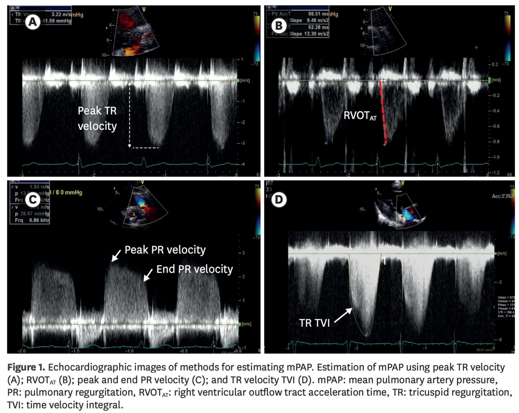
PASP = RVSP
우심실과 폐동맥 사이에 폐색이 없다고 가정하면, 우심실 수축기 압력과 폐동맥 수축기 압력이 같아진다.
따라서 폐동맥 압력은 우심실 수축기 압력을 측정하여 산정할 수 있다.
이것은 삼첨판 역류가 있을 때만 산출이 가능하다.
삼첨판 역류가 우심실과 우심방 사이의 압력차를 만드는데,
이 압력차는 베르누이 공식을 이용하여 최고 속도를 구해 산정할 수 있다.
이 압력차는 우심방 압력도 고려되었으므로 우심실 수축기 압력과 같지 않아
우심방의 압력이 정상인 5mmHg를 더하여 폐동맥 압력을 산출할 수 있다.
RVSP was estimated from the maximal RV–right atrial pressure gradient,
using the modified Bernoulli equation : RVSP = 4 × (maximal TR velocity)² + RAP,
where maximal TR velocity was measured using continuous-wave Doppler.
2. PSAX - level of mitral valve (MV level, MVL) a.k.a. "Fish mouth" view
탐촉자를 PLAX view 위치에서 시계 방향으로 90도 rotation 하여 심첨부로 tilting시켜 승모판을 관찰한다.
해부학적 위치상 흉골 좌연과 늑간 사이에 탐촉자를 위치시킨다.
승모판이 열리고 닫히는 모양을 잘 평가하기 위해서는 좌심실을 완전 구형으로 잘 보이도록 위치시켜야 한다.
이때 승모판 개폐 뿐만 아니라 좌심실 기저부의 벽운동도 같이 관찰할 수 있다.

The base of the mitral valve, or the "fish-mouth” view, will be encountered first, along with the basal segments of the anteroseptal, anterior, anterolateral, inferolateral, inferior and inferoseptal walls of the left ventricle.
우심실의 압력이 정상일 경우 완전 구형으로 좌심실을 관찰할 수 있다.
좌심실의 압력 상승(ex. 폐동맥 혈전증)으로 좌심실에 압력이 가해질 경우, 좌심실의 모양이 D형으로 관찰될 수 있다.
3. PSAX - level of papillary muscle (PML)
심실 기저부면에서 심첨부 쪽으로 탐촉자를 조금 기울여 심실 단면부의 중앙을 관찰한다.
심실 중앙의 단면부 영상은 대표적으로 유두근을 잘 관찰할 수 있어 해부학적 구조를 미리 파악한다.
좌심근이 완전 구형이 되도록 미세하게 탐촉자를 조절하여 좌심 수축기능과 좌심 벽운동을 자세하게 관찰할 수 있다.
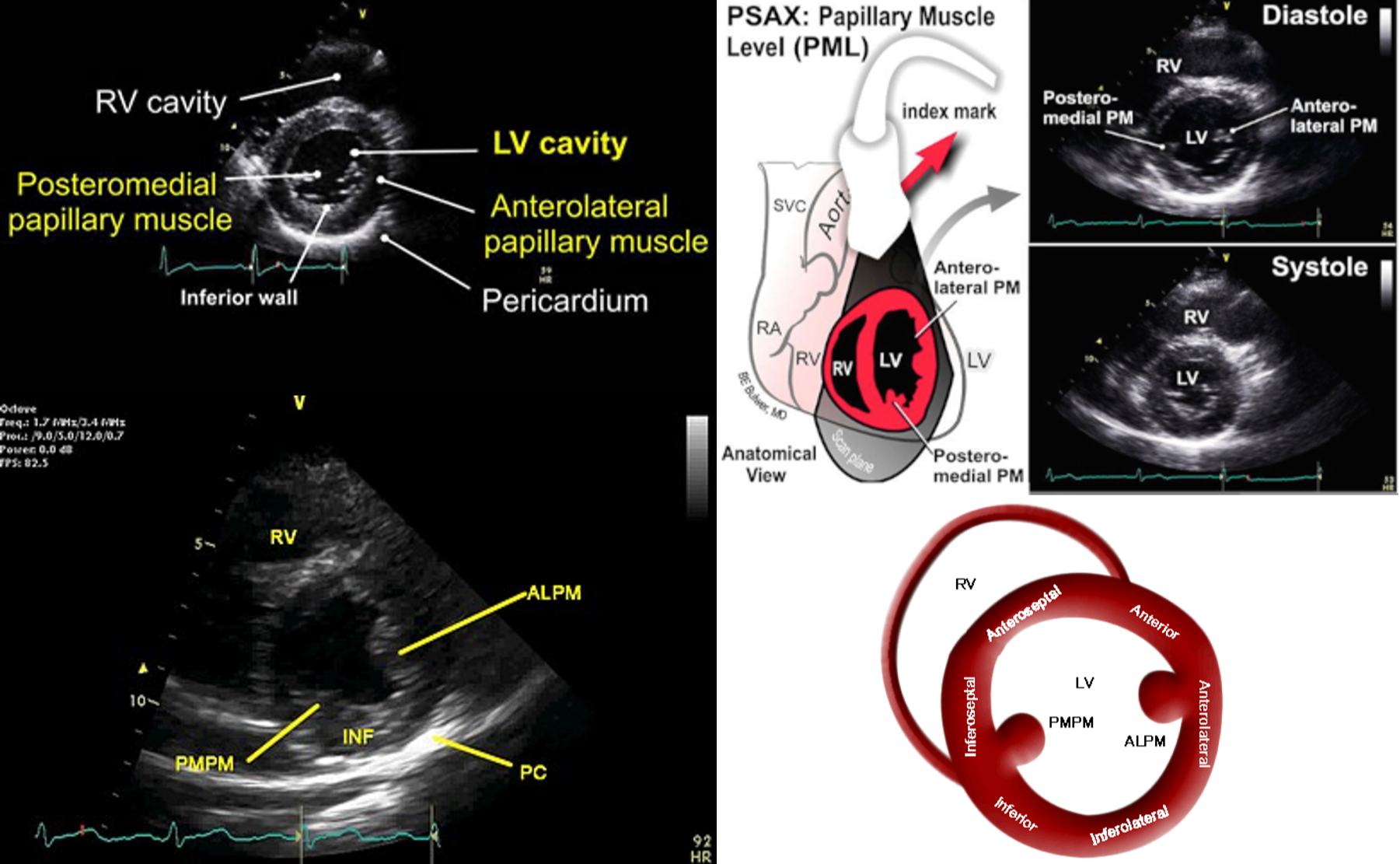
This view is obtained by tilting the tip of the transducer caudal from the mitral valve level.
The middle segments of the anteroseptal, anterior, anterolateral, inferolateral, inferior and inferoseptal walls of the left ventricle are present along with the anterolateral and inferomedial papillary muscles.
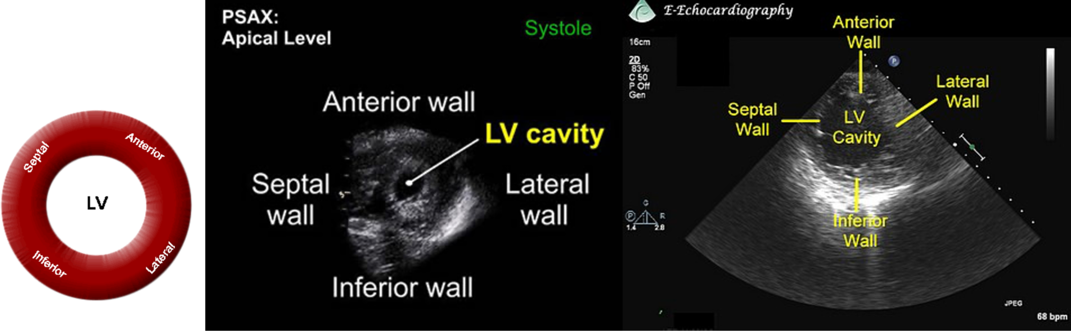
4. PSAX - level of apex (APL)
심실 중간을 관찰할 때보다 심첨부 쪽으로 탐촉자를 더 기울이면 볼 수 있지만
심첨부 단면을 더 정확하게 관찰하려면 심첨부 쪽으로 탐촉자를 이동시킨다.
탐촉자를 늑간 사이에서 겨드랑이 쪽으로 이동시키면 심첨부를 잘 관찰할 수 있다.
탐촉자를 이동할 때 늑간을 잘 눌러서 탐촉자를 이동시키면 뼈에 반사되는 영상을 최소화하여 최적화된 영상을 얻을 수 있다.
'Cardiology' 카테고리의 다른 글
| PDA (동맥관 개존증, Patent ductus arteriosus) (1) | 2023.11.05 |
|---|---|
| ASD (심방중격결손, Atrial septal defect) (0) | 2023.11.05 |
| [심초음파] PLAX or PSLA (parasternal long axis view) (0) | 2023.11.01 |
| 심장초음파의 기본 원리 (1) | 2023.10.30 |
| 심장초음파 (Echo, Echocardiography) (0) | 2023.10.29 |


